Copyright
©2014 Baishideng Publishing Group Inc.
World J Radiol. Jun 28, 2014; 6(6): 274-283
Published online Jun 28, 2014. doi: 10.4329/wjr.v6.i6.274
Published online Jun 28, 2014. doi: 10.4329/wjr.v6.i6.274
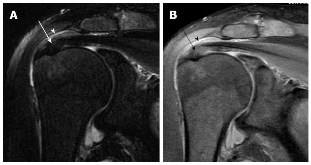
Figure 8 Subacromial impingement.
A: Coronal oblique fat-sat T2-weighted image; B: Coronal oblique fat-sat proton-density-weighted image. Both images show findings associated with subacromial impingement in the form of osteoarthritis of the acromio-clavicular joint, distended subacromial subdeltoid bursa by fluid signal (arrowhead) and focal thickening of the distal supraspinatus tendon with partial irregularity of the bursal surface reported as partial tear (arrow).
- Citation: Tawfik AM, El-Morsy A, Badran MA. Rotator cuff disorders: How to write a surgically relevant magnetic resonance imaging report? World J Radiol 2014; 6(6): 274-283
- URL: https://www.wjgnet.com/1949-8470/full/v6/i6/274.htm
- DOI: https://dx.doi.org/10.4329/wjr.v6.i6.274


