Copyright
©2014 Baishideng Publishing Group Inc.
World J Radiol. Jun 28, 2014; 6(6): 274-283
Published online Jun 28, 2014. doi: 10.4329/wjr.v6.i6.274
Published online Jun 28, 2014. doi: 10.4329/wjr.v6.i6.274
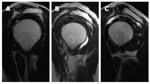
Figure 1 Acromion types.
Sagittal oblique T2-weighted images of different patients showing type I flat acromion (A), type II concave under surface (B) and type III concave under surface with anterior hook (C).
- Citation: Tawfik AM, El-Morsy A, Badran MA. Rotator cuff disorders: How to write a surgically relevant magnetic resonance imaging report? World J Radiol 2014; 6(6): 274-283
- URL: https://www.wjgnet.com/1949-8470/full/v6/i6/274.htm
- DOI: https://dx.doi.org/10.4329/wjr.v6.i6.274


