
















©2014 Baishideng Publishing Group Inc.
World J Radiol. Jun 28, 2014; 6(6): 238-251
Published online Jun 28, 2014. doi: 10.4329/wjr.v6.i6.238
Published online Jun 28, 2014. doi: 10.4329/wjr.v6.i6.238
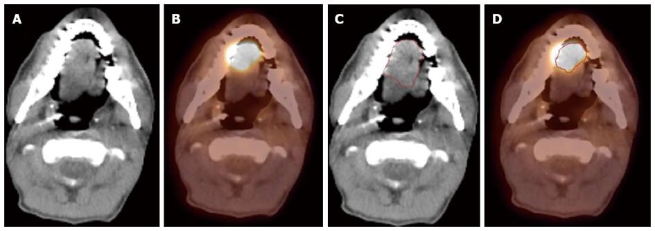
Figure 8 A patient with oral tongue cancer.
The edge of the tumor was not very clear in the computed tomography (CT) image (A), but more obvious in the PET image (B). Gross tumor volume was outlined based on CT scan (C) vs with fluorodeoxyglucose positron emission tomography/CT (D). The volume included is larger with CT alone.
- Citation: Siddiqui F, Yao M. Application of fluorodeoxyglucose positron emission tomography in the management of head and neck cancers. World J Radiol 2014; 6(6): 238-251
- URL: https://www.wjgnet.com/1949-8470/full/v6/i6/238.htm
- DOI: https://dx.doi.org/10.4329/wjr.v6.i6.238