Copyright
©2014 Baishideng Publishing Group Inc.
World J Radiol. Jun 28, 2014; 6(6): 230-237
Published online Jun 28, 2014. doi: 10.4329/wjr.v6.i6.230
Published online Jun 28, 2014. doi: 10.4329/wjr.v6.i6.230
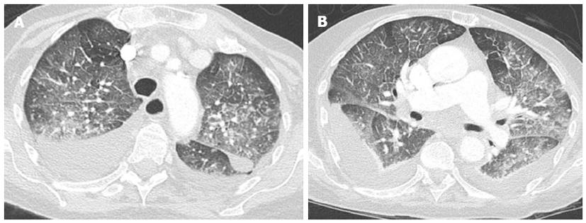
Figure 8 Computed tomography scan through aortic arch and pulmonary arteries planes shows ground-glass opacity with geographic distribution and partial sparing of the lung periphery.
Thickening of interlobular septa and sub-pleural edema and bilateral pleural effusion with passive atelectasis of lower lobes is also present.
- Citation: Cardinale L, Priola AM, Moretti F, Volpicelli G. Effectiveness of chest radiography, lung ultrasound and thoracic computed tomography in the diagnosis of congestive heart failure. World J Radiol 2014; 6(6): 230-237
- URL: https://www.wjgnet.com/1949-8470/full/v6/i6/230.htm
- DOI: https://dx.doi.org/10.4329/wjr.v6.i6.230


