Copyright
©2014 Baishideng Publishing Group Inc.
World J Radiol. Jun 28, 2014; 6(6): 230-237
Published online Jun 28, 2014. doi: 10.4329/wjr.v6.i6.230
Published online Jun 28, 2014. doi: 10.4329/wjr.v6.i6.230
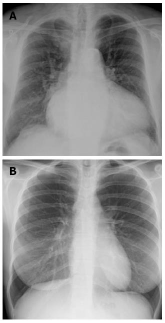
Figure 2 Posterior-anterior chest X-ray demonstrating enlargement of atrial and left ventricles, with redistribution of lung circulation from bases to apex suggestive to pulmonary congestion (A), note the blood vessels are more prominent in the upper lung fields compared to the lung bases, just the opposite of normal (B).
- Citation: Cardinale L, Priola AM, Moretti F, Volpicelli G. Effectiveness of chest radiography, lung ultrasound and thoracic computed tomography in the diagnosis of congestive heart failure. World J Radiol 2014; 6(6): 230-237
- URL: https://www.wjgnet.com/1949-8470/full/v6/i6/230.htm
- DOI: https://dx.doi.org/10.4329/wjr.v6.i6.230


