Copyright
©2014 Baishideng Publishing Group Inc.
World J Radiol. May 28, 2014; 6(5): 148-159
Published online May 28, 2014. doi: 10.4329/wjr.v6.i5.148
Published online May 28, 2014. doi: 10.4329/wjr.v6.i5.148
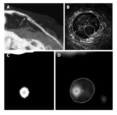
Figure 1 The window settings for the lumen and outer vessel boundary by computed tomography angiography are the same as those for intravascular ultrasound imaging[15].
A: A curved multiplanar reconstructed CTA image reveals a significant stenosis in the left anterior descending artery (arrow); B: An IVUS cross-section reveals a lumen area of 2.1 mm2 and a vessel area of 15.4 mm2; C: The cross-sectional CTA images show the luminal CSA of 2.1 mm2; D: Vessel CSA of 15.4 mm2. CTA: Computed tomography angiography; IVUS: Intravascular ultrasound; CSA: Cross-sectional area.
- Citation: Sato A. Coronary plaque imaging by coronary computed tomography angiography. World J Radiol 2014; 6(5): 148-159
- URL: https://www.wjgnet.com/1949-8470/full/v6/i5/148.htm
- DOI: https://dx.doi.org/10.4329/wjr.v6.i5.148


