Copyright
©2014 Baishideng Publishing Group Inc.
World J Radiol. May 28, 2014; 6(5): 139-147
Published online May 28, 2014. doi: 10.4329/wjr.v6.i5.139
Published online May 28, 2014. doi: 10.4329/wjr.v6.i5.139
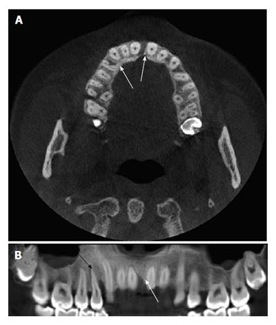
Figure 9 The vertical alveolar bone defects are viewed in cone beam computed tomography images (A, B) (white arrows) and the expansion of periodontal ligament space is also seen in the right maxillary first premolar tooth (B) (black arrow).
- Citation: Acar B, Kamburoğlu K. Use of cone beam computed tomography in periodontology. World J Radiol 2014; 6(5): 139-147
- URL: https://www.wjgnet.com/1949-8470/full/v6/i5/139.htm
- DOI: https://dx.doi.org/10.4329/wjr.v6.i5.139


