Copyright
©2014 Baishideng Publishing Group Co.
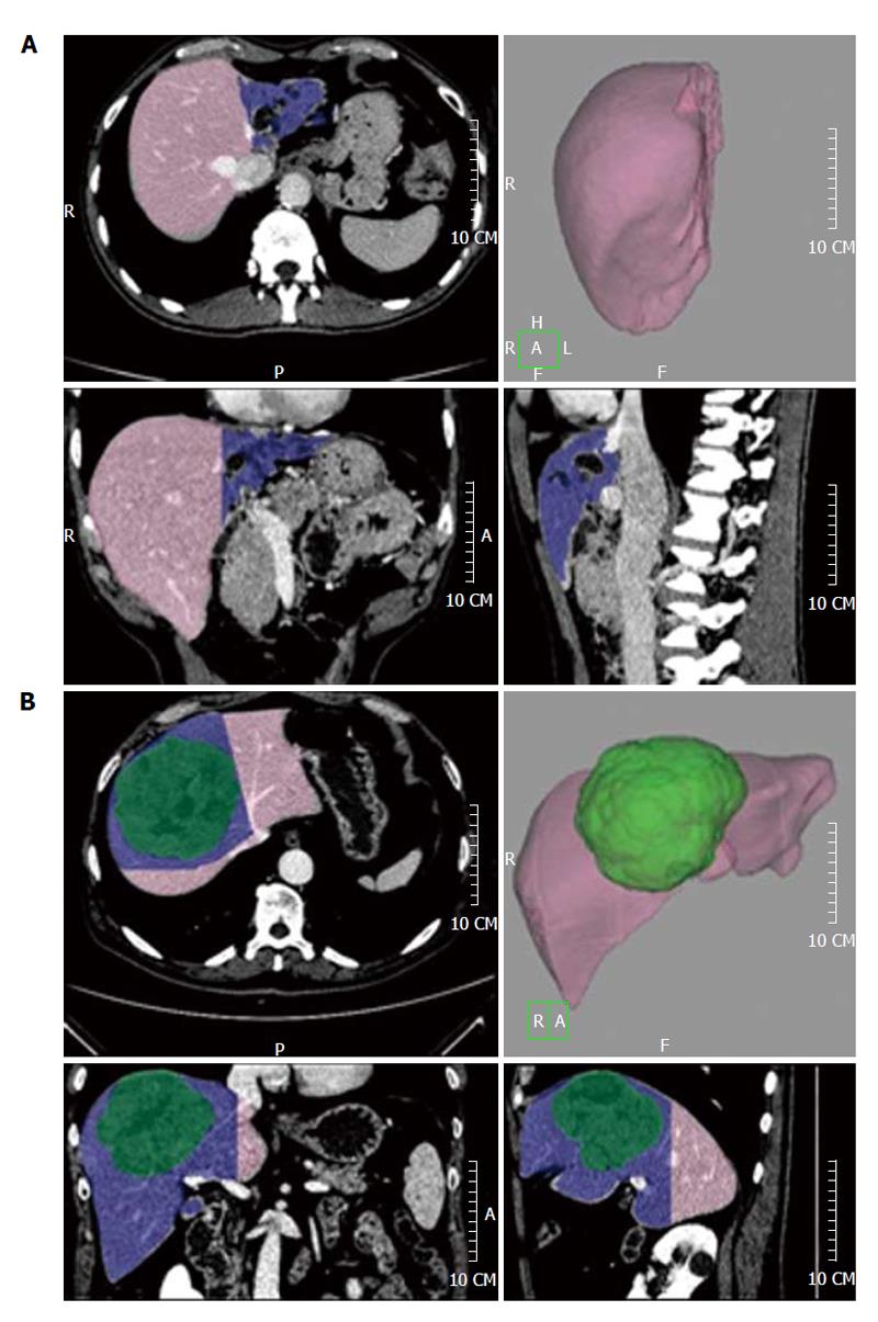
Figure 3 The future liver remnant volume is shown in pink, while the resection volume is shown in blue.
A left hepatectomy for a hilar cholangiocarcinoma involving the left hepatic duct (A) and a mesohepatectomy (resection of liver segments 4-8-5) for a hepatocarcinoma (shown in green, B) are shown.
- Citation: D’Onofrio M, De Robertis R, Demozzi E, Crosara S, Canestrini S, Pozzi Mucelli R. Liver volumetry: Is imaging reliable? Personal experience and review of the literature. World J Radiol 2014; 6(4): 62-71
- URL: https://www.wjgnet.com/1949-8470/full/v6/i4/62.htm
- DOI: https://dx.doi.org/10.4329/wjr.v6.i4.62


