Copyright
©2014 Baishideng Publishing Group Co.
World J Radiol. Apr 28, 2014; 6(4): 125-129
Published online Apr 28, 2014. doi: 10.4329/wjr.v6.i4.125
Published online Apr 28, 2014. doi: 10.4329/wjr.v6.i4.125
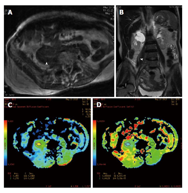
Figure 3 Magnetic resonance imaging.
Axial T1W and coronal T2W equence (A and B) reveals a bulky right psoas muscle (arrowhead) and shows altered signal intensity. On axial diffusion weighted images (C and D) there is e/o restricted diffusion in the involved segment.
- Citation: Basu S, Mahajan A. Psoas muscle metastasis from cervical carcinoma: Correlation and comparison of diagnostic features on FDG-PET/CT and diffusion-weighted MRI. World J Radiol 2014; 6(4): 125-129
- URL: https://www.wjgnet.com/1949-8470/full/v6/i4/125.htm
- DOI: https://dx.doi.org/10.4329/wjr.v6.i4.125


