
















©2014 Baishideng Publishing Group Co.
World J Radiol. Apr 28, 2014; 6(4): 106-115
Published online Apr 28, 2014. doi: 10.4329/wjr.v6.i4.106
Published online Apr 28, 2014. doi: 10.4329/wjr.v6.i4.106
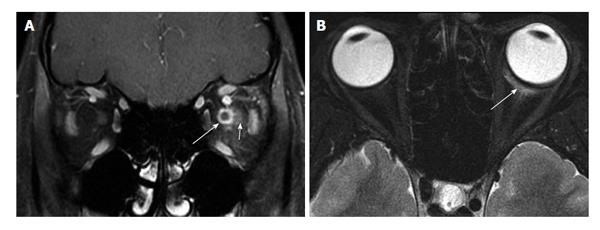
Figure 8 Perineuritic orbital inflammatory disease.
A: Coronal fat-suppressed contrast-enhanced T1 shows circumferential enhancement about the left optic nerve (long arrow), with sparing of the nerve substance. There is also mild infiltration of the surrounding soft tissues (short arrow); B: Axial fat-suppressed T2 shows a small amount of edema about Tenon’s capsule (arrow). This finding, along with clinical history of acute, painful presentation, help distinguish perineuritic pseudotumor from en plaque optic nerve sheath meningioma.
- Citation: Pakdaman MN, Sepahdari AR, Elkhamary SM. Orbital inflammatory disease: Pictorial review and differential diagnosis. World J Radiol 2014; 6(4): 106-115
- URL: https://www.wjgnet.com/1949-8470/full/v6/i4/106.htm
- DOI: https://dx.doi.org/10.4329/wjr.v6.i4.106