Copyright
©2014 Baishideng Publishing Group Co.
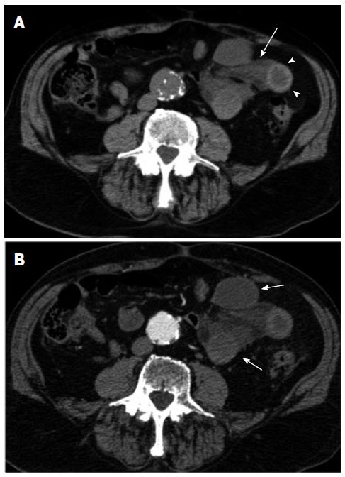
Figure 2 Multi-detector computed tomography.
Unenhanced (A) and contrast-enhanced (B) axial images of the abdomen are shown. In A, a cluster of fluid-filled dilated small bowel loops can be appreciated in the left flank along with the evidence of mesenteric fluid (arrow). A mural hematoma can also be appreciated (arrow-heads). In B lack of enhancement of the intestinal wall of the small bowel loops is depicted as a result of bowel wall ischemia.
- Citation: Camera L, Gennaro AD, Longobardi M, Masone S, Calabrese E, Vecchio WD, Persico G, Salvatore M. A spontaneous strangulated transomental hernia: Prospective and retrospective multi-detector computed tomography findings. World J Radiol 2014; 6(2): 26-30
- URL: https://www.wjgnet.com/1949-8470/full/v6/i2/26.htm
- DOI: https://dx.doi.org/10.4329/wjr.v6.i2.26


