Copyright
©2014 Baishideng Publishing Group Inc.
World J Radiol. Dec 28, 2014; 6(12): 913-918
Published online Dec 28, 2014. doi: 10.4329/wjr.v6.i12.913
Published online Dec 28, 2014. doi: 10.4329/wjr.v6.i12.913
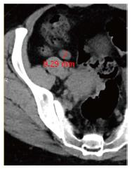
Figure 3 Thirty six-year-old man undergoing evaluation for hematuria.
Axial computed tomography (CT) image showing outer-to-outer wall diameter measures 9.3 mm. A normal, but large diameter appendix with this morphology can be easily mistaken for appendicitis at CT.
- Citation: Yaqoob J, Idris M, Alam MS, Kashif N. Can outer-to-outer diameter be used alone in diagnosing appendicitis on 128-slice MDCT? World J Radiol 2014; 6(12): 913-918
- URL: https://www.wjgnet.com/1949-8470/full/v6/i12/913.htm
- DOI: https://dx.doi.org/10.4329/wjr.v6.i12.913


