Copyright
©2014 Baishideng Publishing Group Inc.
World J Radiol. Nov 28, 2014; 6(11): 886-889
Published online Nov 28, 2014. doi: 10.4329/wjr.v6.i11.886
Published online Nov 28, 2014. doi: 10.4329/wjr.v6.i11.886
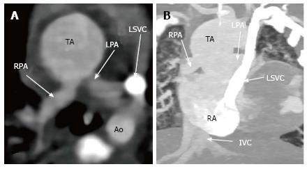
Figure 1 Axial (A) and maximum intensity projection coronal (B) images show a single arterial trunk origin from the right ventricule and left and right branched pulmonary vessels origin from the posterior aspect of trunk.
In addition, persistent left superior vena cava is observed. TA: Truncus arteriosus; LPA: Left pulmonary artery; RPA: Right pulmonary artery; Ao: Descending aorta; LSVC: Left superior vena cava; IVC: Inferior vena cava; RA: Right atrium.
- Citation: Koplay M, Cimen D, Sivri M, Güvenc O, Arslan D, Nayman A, Oran B. Truncus arteriosus: Diagnosis with dual-source computed tomography angiography and low radiation dose. World J Radiol 2014; 6(11): 886-889
- URL: https://www.wjgnet.com/1949-8470/full/v6/i11/886.htm
- DOI: https://dx.doi.org/10.4329/wjr.v6.i11.886


