
















©2014 Baishideng Publishing Group Inc.
World J Radiol. Oct 28, 2014; 6(10): 808-825
Published online Oct 28, 2014. doi: 10.4329/wjr.v6.i10.808
Published online Oct 28, 2014. doi: 10.4329/wjr.v6.i10.808
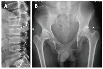
Figure 2 Spondyloepiphyseal dysplasia tarda.
Lateral radiograph of lumbar spine (A) shows characteristic posterior hump (arrow). Radiograph of pelvis (B) shows bilateral flattened femoral heads, short necks and premature degenerative changes (arrow).
- Citation: Panda A, Gamanagatti S, Jana M, Gupta AK. Skeletal dysplasias: A radiographic approach and review of common non-lethal skeletal dysplasias. World J Radiol 2014; 6(10): 808-825
- URL: https://www.wjgnet.com/1949-8470/full/v6/i10/808.htm
- DOI: https://dx.doi.org/10.4329/wjr.v6.i10.808