Copyright
©2014 Baishideng Publishing Group Inc.
World J Radiol. Oct 28, 2014; 6(10): 741-755
Published online Oct 28, 2014. doi: 10.4329/wjr.v6.i10.741
Published online Oct 28, 2014. doi: 10.4329/wjr.v6.i10.741
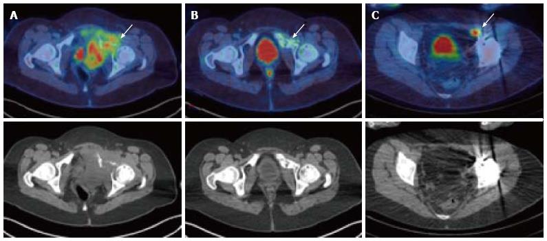
Figure 3 A 17-year-old female with Ewing’s sarcoma involving the left superior pubic ramus.
Staging PET/CT showed extensive disease (A, arrow) with bone destruction and a large FDG-avid pelvic mass. Following chemotherapy, at 5 mo after diagnosis, there was an excellent metabolic response with minimal residual FDG uptake (B, arrow). Patient underwent surgical resection and extracorporeal radiotherapy to the bone. Sixteen months after completion of treatment, surveillance PET/CT demonstrated recurrence in a left external iliac lymph node (C) which was not detectable on the CT or MRI due to marked metal artefact. PET/CT: Positron emission tomography/computed tomography; FDG: 2-deoxy-2-(18F)fluoro-D-glucose; MRI: Magnetic resonance imaging.
- Citation: Freebody J, Wegner EA, Rossleigh MA. 2-deoxy-2-(18F)fluoro-D-glucose positron emission tomography/computed tomography imaging in paediatric oncology. World J Radiol 2014; 6(10): 741-755
- URL: https://www.wjgnet.com/1949-8470/full/v6/i10/741.htm
- DOI: https://dx.doi.org/10.4329/wjr.v6.i10.741


