
















©2013 Baishideng Publishing Group Co.
World J Radiol. Sep 28, 2013; 5(9): 345-348
Published online Sep 28, 2013. doi: 10.4329/wjr.v5.i9.345
Published online Sep 28, 2013. doi: 10.4329/wjr.v5.i9.345
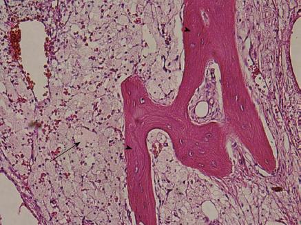
Figure 4 Hematoxylin and eosin stained image (× 125 magnification).
The biopsy specimen demonstrating foamy lipidic histiocytic infiltration (arrow) of marrow; Trabeculae are noted (arrowheads).
- Citation: Ali S, Fedenko A, Syed AB, Matcuk G, Patel D, Gottsegen C, White E. Bilateral primary xanthoma of the humeri with pathologic fractures: A case report. World J Radiol 2013; 5(9): 345-348
- URL: https://www.wjgnet.com/1949-8470/full/v5/i9/345.htm
- DOI: https://dx.doi.org/10.4329/wjr.v5.i9.345