Copyright
©2013 Baishideng Publishing Group Co.
World J Radiol. Aug 28, 2013; 5(8): 328-333
Published online Aug 28, 2013. doi: 10.4329/wjr.v5.i8.328
Published online Aug 28, 2013. doi: 10.4329/wjr.v5.i8.328
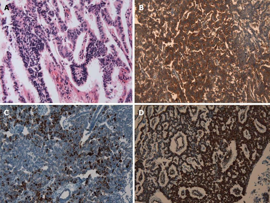
Figure 4 Carcinoid tumors of the kidney are composed of nests and cords of neuroendocrine cells that are noticeable under microscopy.
Small- to intermediate-sized tumor cells have uniformly round nuclei with net formation, abundant cytoplasm, and prominent rosette-like structures (A, HE stain, ×200). The neoplasm had low mitotic activity (1/10 HPF). Immunohistochemical studies were performed, and the tumor cells tested strongly positive for the neuroendocrine markers (×200), CD99 (B), chromogranin (C), and vimentin (D). The cells were negative for cytokeratin, the marker for renal cell carcinoma (not shown). Microscopic and immunohistochemical findings were consistent with a well-differentiated neuroendocrine tumor, so called carcinoid tumor.
- Citation: Yoon JH. Primary renal carcinoid tumor: A rare cystic renal neoplasm. World J Radiol 2013; 5(8): 328-333
- URL: https://www.wjgnet.com/1949-8470/full/v5/i8/328.htm
- DOI: https://dx.doi.org/10.4329/wjr.v5.i8.328


