Copyright
©2013 Baishideng Publishing Group Co.
World J Radiol. Aug 28, 2013; 5(8): 328-333
Published online Aug 28, 2013. doi: 10.4329/wjr.v5.i8.328
Published online Aug 28, 2013. doi: 10.4329/wjr.v5.i8.328
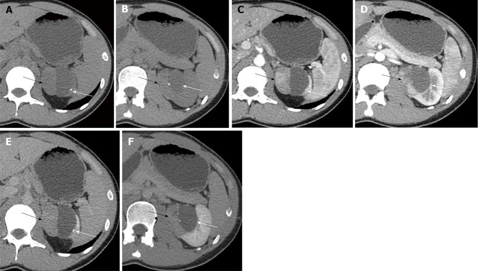
Figure 1 Axial images of the dynamic abdominal computer tomography scans show a well-circumscribed, bilobed renal mass in the left kidney that is composed of a solid part and a cystic part containing peripheral wall and septal calcifications.
A, B: Pre-contrast images; C, D: Corticomedullary phase images; E, F: Nephrographic phase images. The lesion has significant enhancement in the right half of the solid portion during the corticomedullary phase and enhancement washout during the nephrographic phase (black arrow). A non-enhancing cystic portion is evident in the left half (white arrow).
- Citation: Yoon JH. Primary renal carcinoid tumor: A rare cystic renal neoplasm. World J Radiol 2013; 5(8): 328-333
- URL: https://www.wjgnet.com/1949-8470/full/v5/i8/328.htm
- DOI: https://dx.doi.org/10.4329/wjr.v5.i8.328


