Copyright
©2013 Baishideng Publishing Group Co.
World J Radiol. Aug 28, 2013; 5(8): 275-284
Published online Aug 28, 2013. doi: 10.4329/wjr.v5.i8.275
Published online Aug 28, 2013. doi: 10.4329/wjr.v5.i8.275
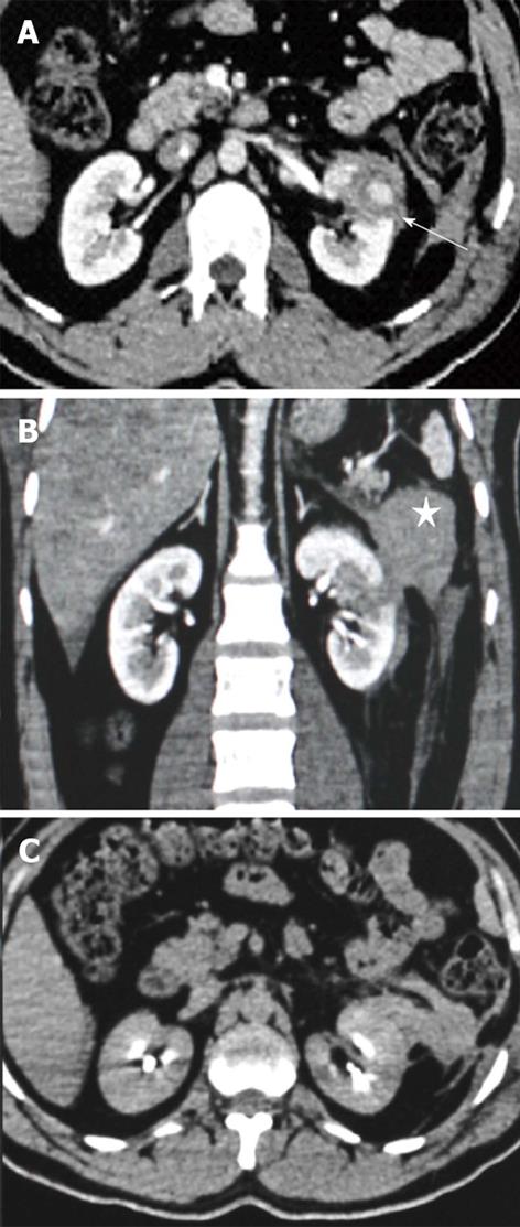
Figure 4 Grade III injury.
Contrast enhanced computed tomography (CECT) images of a 27-year-old male patient who had a stab injury in the left flank. A: Axial section showing a deep laceration (arrow) reaching up to the hilum; B: Coronal section showing the deep laceration at mid pole with disruption of the pararenal fascia and a large hematoma (star) lateral to it; C: Axial delayed image in excretory phase showed no excreted contrast extravasation.
- Citation: Dayal M, Gamanagatti S, Kumar A. Imaging in renal trauma. World J Radiol 2013; 5(8): 275-284
- URL: https://www.wjgnet.com/1949-8470/full/v5/i8/275.htm
- DOI: https://dx.doi.org/10.4329/wjr.v5.i8.275


