Copyright
©2013 Baishideng Publishing Group Co.
World J Radiol. Jun 28, 2013; 5(6): 229-240
Published online Jun 28, 2013. doi: 10.4329/wjr.v5.i6.229
Published online Jun 28, 2013. doi: 10.4329/wjr.v5.i6.229
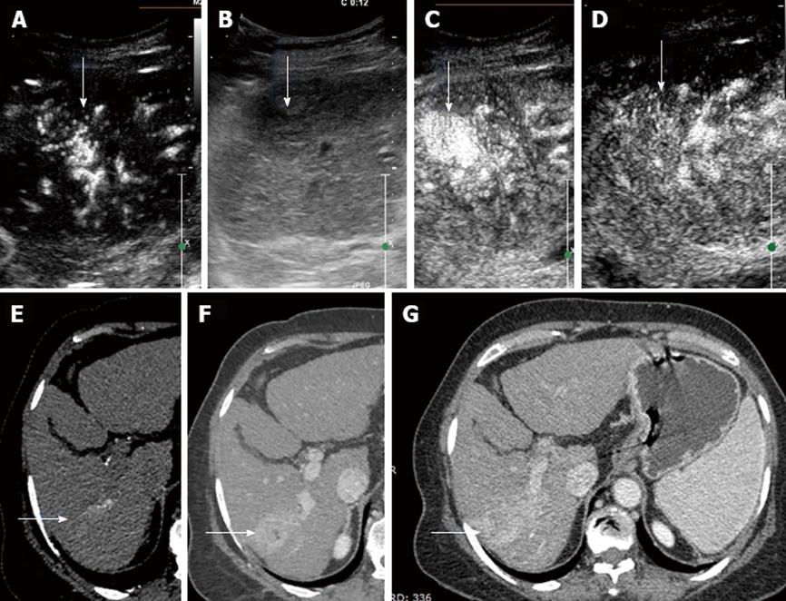
Figure 4 Hepatocellular carcinoma with classical enhancement pattern on contrast enhanced ultrasound in a 50-year-old patient with hepatitis B infection, detected to have a mass on routine screening.
A: Early arterial enhancement of the mass on contrast enhanced ultrasound (CEUS) (arrow); B: B mode synchronous image of the mass lesion (arrow); C: Rapid contrast enhancement of the lesion on CEUS (arrow); D: Early washout of contrast within the lesion (arrow) on CEUS; E: Arterial phase enhancement of the lesion on triple phase computer tomography (CT) scan (arrow); F: Portal venous phase on dynamic CT showing lesion enhancement (arrow); G: Equilibrium Phase on CT showing lesion contrast washout (arrow).
- Citation: Laroia ST, Bawa SS, Jain D, Mukund A, Sarin S. Contrast ultrasound in hepatocellular carcinoma at a tertiary liver center: First Indian experience. World J Radiol 2013; 5(6): 229-240
- URL: https://www.wjgnet.com/1949-8470/full/v5/i6/229.htm
- DOI: https://dx.doi.org/10.4329/wjr.v5.i6.229


