Copyright
©2013 Baishideng.
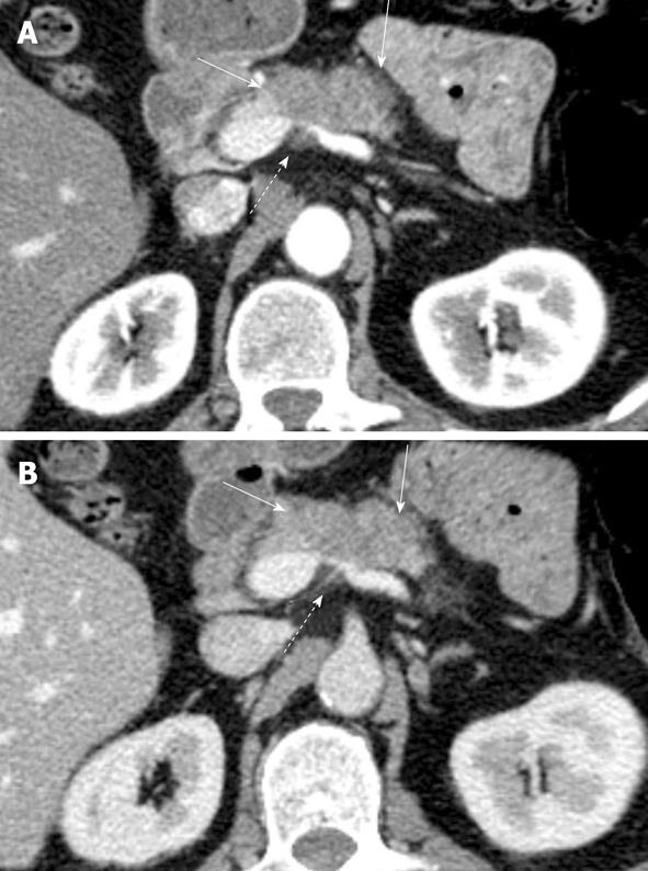
Figure 1 Axial images from dual phase pancreas protocol contrast enhanced computed tomography examination in patient with history of pancreatic cancer shows (A) better demarcation of tumor (solid arrows) on the pancreatic parenchymal phase than on the (B) portal venous phase.
There is marked narrowing of the splenoportal confluence (dashed arrows) secondary to encasement.
- Citation: Tamm EP, Bhosale PR, Vikram R, de Almeida Marcal LP, Balachandran A. Imaging of pancreatic ductal adenocarcinoma: State of the art. World J Radiol 2013; 5(3): 98-105
- URL: https://www.wjgnet.com/1949-8470/full/v5/i3/98.htm
- DOI: https://dx.doi.org/10.4329/wjr.v5.i3.98


