Copyright
©2013 Baishideng.
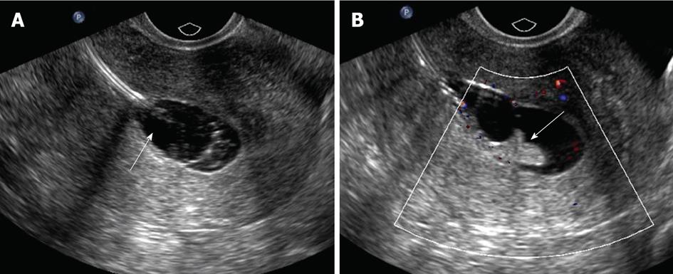
Figure 2 Common pitfalls during sonohysterography.
A: Longitudinal ultrasound image showing balloon hyperinflation during sonohysterography (arrow), which displaces and obscures an endometrial polyp; B: After the balloon is deflated to an adequate level, the echogenic endometrial polyp becomes evident (arrow).
- Citation: Yang T, Pandya A, Marcal L, Bude RO, Platt JF, Bedi DG, Elsayes KM. Sonohysterography: Principles, technique and role in diagnosis of endometrial pathology. World J Radiol 2013; 5(3): 81-87
- URL: https://www.wjgnet.com/1949-8470/full/v5/i3/81.htm
- DOI: https://dx.doi.org/10.4329/wjr.v5.i3.81


