Copyright
©2013 Baishideng.
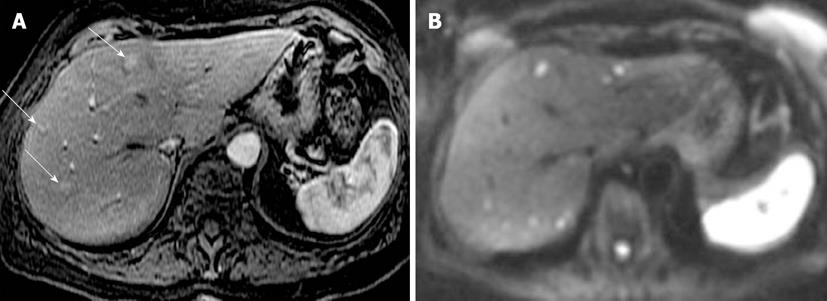
Figure 2 Neuroendocrine metastases to liver.
A: Axial arterial phase post contrast T1-weighted (T1W) gradient recalled-echo; B: Diffusion-weighted image (DWI). Only 3 enhancing metastatic lesions are seen on the post contrast T1W image. DWI increases conspicuity of these lesions and also shows many more bilobar high signal metastatic lesions with impeded diffusion.
- Citation: Morani AC, Elsayes KM, Liu PS, Weadock WJ, Szklaruk J, Dillman JR, Khan A, Chenevert TL, Hussain HK. Abdominal applications of diffusion-weighted magnetic resonance imaging: Where do we stand. World J Radiol 2013; 5(3): 68-80
- URL: https://www.wjgnet.com/1949-8470/full/v5/i3/68.htm
- DOI: https://dx.doi.org/10.4329/wjr.v5.i3.68


