
















©2013 Baishideng.
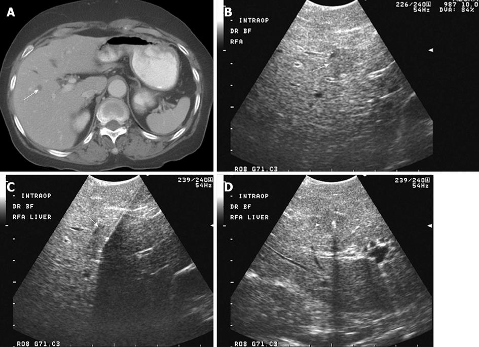
Figure 1 A 79-year-old male with metastatic colon with rising carcinoembryonic antigen and new hypodense lesion in the right liver on computed tomography examination.
A: Axial contrast-enhanced computed tomography shows a small hypodense lesion at the junction of segments VIII and V, concerning for metastasis; B: Intraoperative transverse sonogram identifies a solid slightly hypoechoic lesion in the right liver, at the junction of segments VIII-V, compatible with a metastasis; C, D: Longitudinal and transverse intraoperative ultrasound image shows the position of a radiofrequency ablation needle, with its in appropriate location within the center of the lesion. Intraoperative ultrasound is extremely useful to precise localize hepatic lesions and guide therapeutic interventions.
- Citation: Marcal LP, Patnana M, Bhosale P, Bedi DG. Intraoperative abdominal ultrasound in oncologic imaging. World J Radiol 2013; 5(3): 51-60
- URL: https://www.wjgnet.com/1949-8470/full/v5/i3/51.htm
- DOI: https://dx.doi.org/10.4329/wjr.v5.i3.51