Copyright
©2013 Baishideng.
World J Radiol. Mar 28, 2013; 5(3): 126-142
Published online Mar 28, 2013. doi: 10.4329/wjr.v5.i3.126
Published online Mar 28, 2013. doi: 10.4329/wjr.v5.i3.126
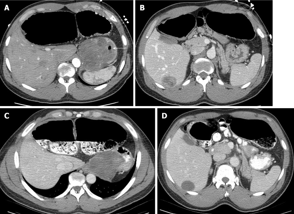
Figure 6 A computed tomography image of a 64-year-old male with metastatic gastro intestinal tumors prior to the imatinib shows a large heterogeneously enhanced gastric mass compatible with gastric gastro intestinal tumors (A) and a segment 6 hepatic metastasis (B).
The primary tumor and hepatic metastasis showed decreases in tumor size and became homogeneous in internal appearance after the targeted therapy (C, D). Note the stomach air bubble (arrow in A).
- Citation: Peungjesada S, Chuang HH, Prasad SR, Choi H, Loyer EM, Bronstein Y. Evaluation of cancer treatment in the abdomen: Trends and advances. World J Radiol 2013; 5(3): 126-142
- URL: https://www.wjgnet.com/1949-8470/full/v5/i3/126.htm
- DOI: https://dx.doi.org/10.4329/wjr.v5.i3.126


