Copyright
©2013 Baishideng.
World J Radiol. Mar 28, 2013; 5(3): 126-142
Published online Mar 28, 2013. doi: 10.4329/wjr.v5.i3.126
Published online Mar 28, 2013. doi: 10.4329/wjr.v5.i3.126
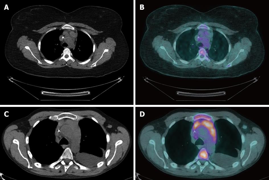
Figure 5 A computed tomography image of a 22-year-old female with Hodgkin’s lymphoma after 4 cycles of chemotherapy demonstrated residual mediastinal lymphadenopathy (A) that is not metabolically active, as seen on positron emission tomography/computed tomography fusion images, and consistent with post-therapy fibrosis (B).
A computed tomography image of a 50-year-old male with diffuse large B-cell lymphoma after 6 cycles of chemotherapy showed a residual mediastinal abnormality that represents viable tumor as shown by the abnormal actiivty on the positron emission tomography/computed tomography fusion image. Also note left pleural effusion and bone marrow activation (D).
- Citation: Peungjesada S, Chuang HH, Prasad SR, Choi H, Loyer EM, Bronstein Y. Evaluation of cancer treatment in the abdomen: Trends and advances. World J Radiol 2013; 5(3): 126-142
- URL: https://www.wjgnet.com/1949-8470/full/v5/i3/126.htm
- DOI: https://dx.doi.org/10.4329/wjr.v5.i3.126


