Copyright
©2013 Baishideng.
World J Radiol. Mar 28, 2013; 5(3): 126-142
Published online Mar 28, 2013. doi: 10.4329/wjr.v5.i3.126
Published online Mar 28, 2013. doi: 10.4329/wjr.v5.i3.126
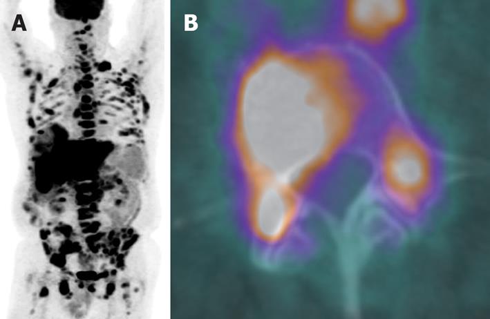
Figure 4 A whole body positron emission tomography image of a 67-year-old male with marginal zone lymphoma showed extensive disease involving lymph nodes above and below diaphragm, osseous structures and liver (A).
The bone marrow biopsy was negative but the direct biopsy from L4, which was positive on the positron emission tomography/computed tomography fusion image (B), confirmed bone marrow involvement.
- Citation: Peungjesada S, Chuang HH, Prasad SR, Choi H, Loyer EM, Bronstein Y. Evaluation of cancer treatment in the abdomen: Trends and advances. World J Radiol 2013; 5(3): 126-142
- URL: https://www.wjgnet.com/1949-8470/full/v5/i3/126.htm
- DOI: https://dx.doi.org/10.4329/wjr.v5.i3.126


