
















©2013 Baishideng.
World J Radiol. Mar 28, 2013; 5(3): 113-125
Published online Mar 28, 2013. doi: 10.4329/wjr.v5.i3.113
Published online Mar 28, 2013. doi: 10.4329/wjr.v5.i3.113
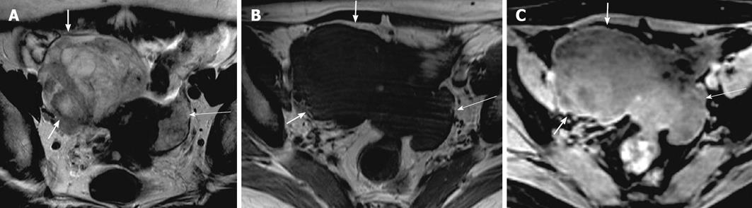
Figure 14 Mucinous cystadenocarcinoma.
A 47-year-old woman with mobile pelvic mass. Axial T2 weighted image (A), axial pre contrast and post contrast T1 weighted images (B and C) demonstrating a right ovarian large heterogenous complex mass, which has a relatively high signal intensity on T2 weighted image (thick arrow) and low signal intensity on pre contrast T1 weighted image with a significant post contrast heterogenous enhancement. Another smaller lesion with similar characteristic is noted in the left adnexa (thin long arrow), representing bilateral surgically proven mucinous cystadenocarcinoma.
- Citation: Wasnik AP, Menias CO, Platt JF, Lalchandani UR, Bedi DG, Elsayes KM. Multimodality imaging of ovarian cystic lesions: Review with an imaging based algorithmic approach. World J Radiol 2013; 5(3): 113-125
- URL: https://www.wjgnet.com/1949-8470/full/v5/i3/113.htm
- DOI: https://dx.doi.org/10.4329/wjr.v5.i3.113