Copyright
©2013 Baishideng.
World J Radiol. Mar 28, 2013; 5(3): 113-125
Published online Mar 28, 2013. doi: 10.4329/wjr.v5.i3.113
Published online Mar 28, 2013. doi: 10.4329/wjr.v5.i3.113
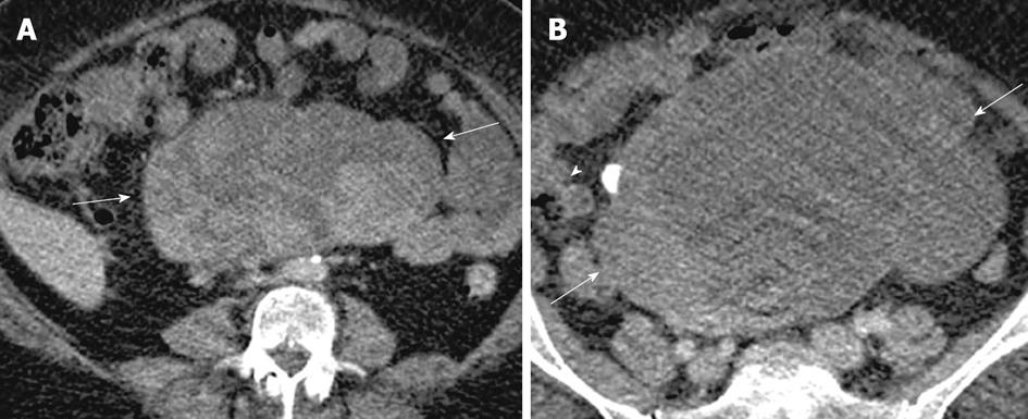
Figure 12 Clear cell carcinoma.
A 49-year-old woman with abdominopelvic mass. Contrast enhanced computed tomography images of the lower abdomen and pelvis (A and B), showing a large lobulated complex cystic mass with enhancing nodular and solid component (arrows) and also a calcific focus (arrowhead).
- Citation: Wasnik AP, Menias CO, Platt JF, Lalchandani UR, Bedi DG, Elsayes KM. Multimodality imaging of ovarian cystic lesions: Review with an imaging based algorithmic approach. World J Radiol 2013; 5(3): 113-125
- URL: https://www.wjgnet.com/1949-8470/full/v5/i3/113.htm
- DOI: https://dx.doi.org/10.4329/wjr.v5.i3.113


