
















©2013 Baishideng Publishing Group Co.
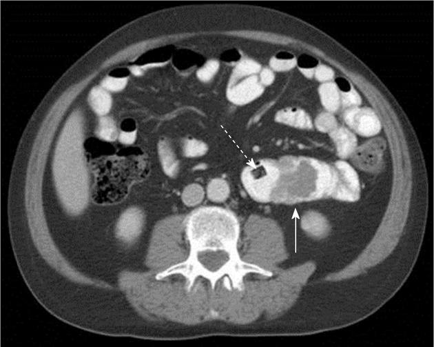
Figure 3 Small bowel metastasis.
Axial section from a contrast enhanced computed tomography in this 54-year-old man with metastatic melanoma shows a partially obstructing endolumenal mass (white arrow) in the jejunum. This patient presented with recurrent gastrointestinal bleeding. Note the video endoscopy capsule that failed to pass the metastatic implant (dashed arrow).
- Citation: Trout AT, Rabinowitz RS, Platt JF, Elsayes KM. Melanoma metastases in the abdomen and pelvis: Frequency and patterns of spread. World J Radiol 2013; 5(2): 25-32
- URL: https://www.wjgnet.com/1949-8470/full/v5/i2/25.htm
- DOI: https://dx.doi.org/10.4329/wjr.v5.i2.25