Copyright
©2013 Baishideng Publishing Group Co.
World J Radiol. Dec 28, 2013; 5(12): 460-467
Published online Dec 28, 2013. doi: 10.4329/wjr.v5.i12.460
Published online Dec 28, 2013. doi: 10.4329/wjr.v5.i12.460
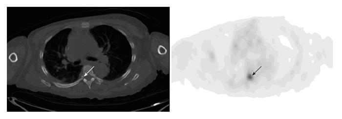
Figure 12 Osseous uptake.
A 55-year-old woman with history of metastatic breast cancer had fluorodeoxyglucose positron emission tomography-computer tomography (PET-CT) for restaging. Axial PET image of the chest shows focal uptake at the right pedicle of the T6 (arrow). There is no visible corresponding bone lesion on the integrated CT (arrow). Repeat PET-CT three months after shows multiple bone metastases including worsening uptake in the right-sided T6.
- Citation: Liu Y. Fluorodeoxyglucose uptake in absence of CT abnormality on PET-CT: What is it? World J Radiol 2013; 5(12): 460-467
- URL: https://www.wjgnet.com/1949-8470/full/v5/i12/460.htm
- DOI: https://dx.doi.org/10.4329/wjr.v5.i12.460


