Copyright
©2013 Baishideng Publishing Group Co.
World J Radiol. Dec 28, 2013; 5(12): 460-467
Published online Dec 28, 2013. doi: 10.4329/wjr.v5.i12.460
Published online Dec 28, 2013. doi: 10.4329/wjr.v5.i12.460
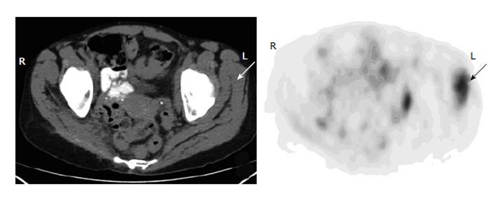
Figure 9 Muscular uptake.
A 45-year-old woman with history of leiomyosarcoma had fluorodeoxyglucose positron emission tomography-computer tomography (PET-CT) for restaging. Axial PET-CT images of the pelvis show intense focal muscular uptake in the left gluteus maximus, gluteus minimus and obturator intermus, with no corresponding lesions on the CT (arrows). Two subsequent magnetic resonance images were unremarkable of the muscles.
- Citation: Liu Y. Fluorodeoxyglucose uptake in absence of CT abnormality on PET-CT: What is it? World J Radiol 2013; 5(12): 460-467
- URL: https://www.wjgnet.com/1949-8470/full/v5/i12/460.htm
- DOI: https://dx.doi.org/10.4329/wjr.v5.i12.460


