Copyright
©2013 Baishideng Publishing Group Co.
World J Radiol. Dec 28, 2013; 5(12): 455-459
Published online Dec 28, 2013. doi: 10.4329/wjr.v5.i12.455
Published online Dec 28, 2013. doi: 10.4329/wjr.v5.i12.455
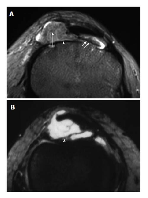
Figure 10 Localized nodular synovitis (solitary PVNS).
A: Axial T2-weighted MR image of the knee shows a nodular mass (arrowhead), with a long pedicle (double short arrows) attaching the mass to the adjacent synovium, involving the infrapatellar fat pad. Note small circular foci of low signal intensity (arrow), corresponding to deposition of hemosiderin. B: Gadolinium-enhanced T1-weighted image with fat saturation shows obvious enhancement of the lesion (arrow) caused by capillary proliferation. (Photo courtesy of Dr. Guo-Shu Huang). MR: Magnetic resonance.
- Citation: Chan WP. Magnetic resonance imaging of soft-tissue tumors of the extremities: A practical approach. World J Radiol 2013; 5(12): 455-459
- URL: https://www.wjgnet.com/1949-8470/full/v5/i12/455.htm
- DOI: https://dx.doi.org/10.4329/wjr.v5.i12.455


