Copyright
©2013 Baishideng Publishing Group Co.
World J Radiol. Dec 28, 2013; 5(12): 455-459
Published online Dec 28, 2013. doi: 10.4329/wjr.v5.i12.455
Published online Dec 28, 2013. doi: 10.4329/wjr.v5.i12.455
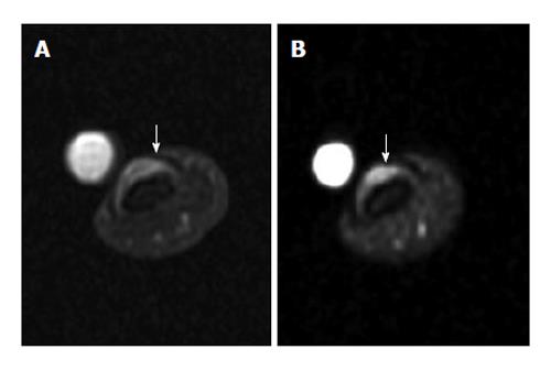
Figure 9 Subungual glomus tumor.
A: Axial proton-density-weighted MR image of the left thumb shows a small subungual tumor (arrow); B: Fat-saturated gadolinium-enhanced T1-weighted image shows obvious enhancement of the tumor (arrow). MR: Magnetic resonance.
- Citation: Chan WP. Magnetic resonance imaging of soft-tissue tumors of the extremities: A practical approach. World J Radiol 2013; 5(12): 455-459
- URL: https://www.wjgnet.com/1949-8470/full/v5/i12/455.htm
- DOI: https://dx.doi.org/10.4329/wjr.v5.i12.455


