
















©2013 Baishideng Publishing Group Co.
World J Radiol. Nov 28, 2013; 5(11): 436-445
Published online Nov 28, 2013. doi: 10.4329/wjr.v5.i11.436
Published online Nov 28, 2013. doi: 10.4329/wjr.v5.i11.436
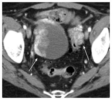
Figure 16 A 60-year-old female status post left radical nephrectomy with liver and bony metastases and with bilateral ovarian metastases.
Axial CT image in arterial phase shows a large cystic and solid mass seen in right ovary and solid hypervascular mass in left ovary. The solid components of the cystic mass are hypervascular on arterial phase.
- Citation: Coquia SF, Johnson PT, Ahmed S, Fishman EK. MDCT imaging following nephrectomy for renal cell carcinoma: Protocol optimization and patterns of tumor recurrence. World J Radiol 2013; 5(11): 436-445
- URL: https://www.wjgnet.com/1949-8470/full/v5/i11/436.htm
- DOI: https://dx.doi.org/10.4329/wjr.v5.i11.436