Copyright
©2013 Baishideng Publishing Group Co.
World J Radiol. Nov 28, 2013; 5(11): 430-435
Published online Nov 28, 2013. doi: 10.4329/wjr.v5.i11.430
Published online Nov 28, 2013. doi: 10.4329/wjr.v5.i11.430
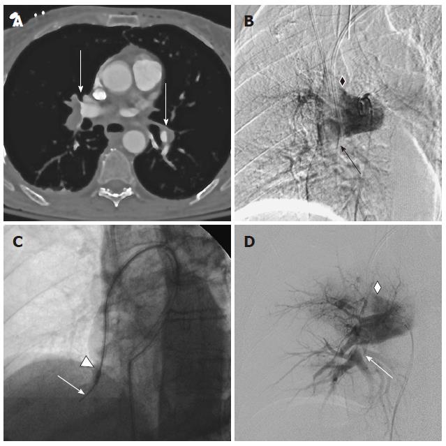
Figure 2 Treatment of massive pulmonary embolism with an aspiration catheter.
A: Axial contrast enhanced computed tomography (CT) at time of diagnosis: Extensive bilateral PE with complete occlusion of the right pulmonary artery with thrombus in the ascending branch and the inferior trunk, partial obstruction of the left upper lobe branch (arrows). Left subcutaneous emphysema from vigorous resuscitation is noted; B: Digital Subtraction Angiography of the right pulmonary artery (RAO): Angiographic findings confirm complete obstruction of the ascending branch (diamond) and the interlobar artery of the right pulmonary artery (arrow); C: Angiographic imaging of pulmonary artery embolus aspiration. A 10 F aspiration catheter (Pronto .035”) is advanced into the thrombus over a guidewire (arrow). Radio-opaque marking of the catheter tip enables controlled positioning of the aspiration side hole just proximal to the tip in the thrombus (Triangle); D: Digital Subtraction Angiography of the right pulmonary artery (RAO): After aspiration maneuvers, selective angiography in the interlobar artery confirms reestablished contrast flow in the interlobar artery with remaining thrombus (arrow), reflux in the ascending branch (diamond) is visualized.
- Citation: Heberlein WE, Meek ME, Saleh O, Meek JC, Lensing SY, Culp WC. New generation aspiration catheter: Feasibility in the treatment of pulmonary embolism. World J Radiol 2013; 5(11): 430-435
- URL: https://www.wjgnet.com/1949-8470/full/v5/i11/430.htm
- DOI: https://dx.doi.org/10.4329/wjr.v5.i11.430


