Copyright
©2012 Baishideng Publishing Group Co.
World J Radiol. Sep 28, 2012; 4(9): 413-417
Published online Sep 28, 2012. doi: 10.4329/wjr.v4.i9.413
Published online Sep 28, 2012. doi: 10.4329/wjr.v4.i9.413
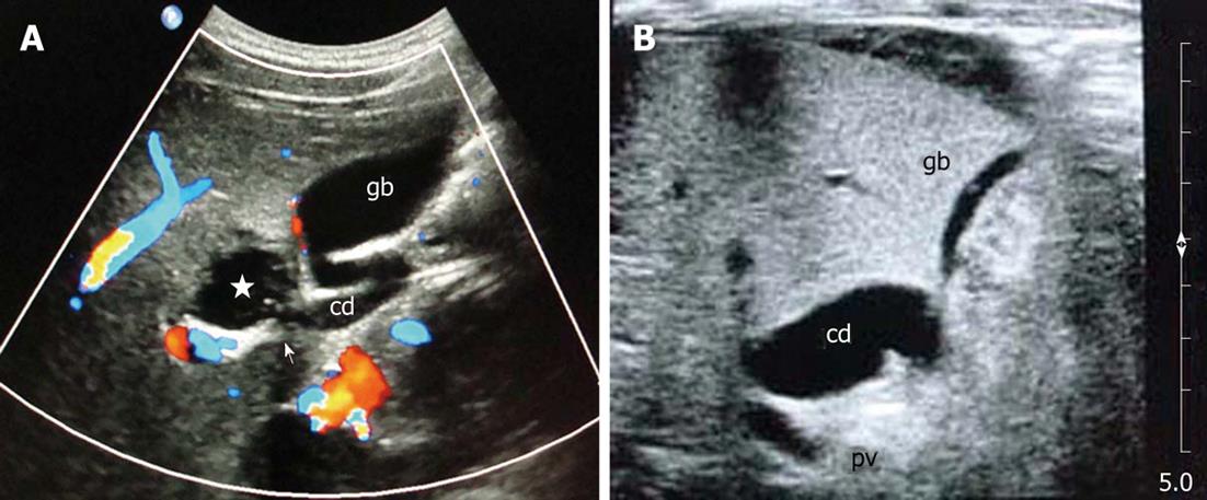
Figure 2 Ultrasonography.
A: Focal saccular dilatation of the cystic duct (star), arrow points to communication of the cyst with cystic duct; B: Mild fusiform dilatation of the cystic duct. gb: Gallbladder; cd: Cystic duct.
- Citation: Maheshwari P. Cystic malformation of cystic duct: 10 cases and review of literature. World J Radiol 2012; 4(9): 413-417
- URL: https://www.wjgnet.com/1949-8470/full/v4/i9/413.htm
- DOI: https://dx.doi.org/10.4329/wjr.v4.i9.413


