Copyright
©2012 Baishideng Publishing Group Co.
World J Radiol. May 28, 2012; 4(5): 186-192
Published online May 28, 2012. doi: 10.4329/wjr.v4.i5.186
Published online May 28, 2012. doi: 10.4329/wjr.v4.i5.186
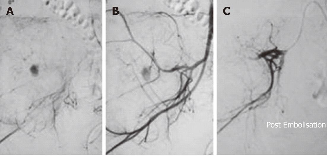
Figure 4 Right iliac giant cell tumor Embolisation in a 25-year-old female.
A: Twenty-five-year old female with a large lytic expansile lesion involving the right iliac wing confirmed to be a giant cell tumor on histopathology. Internal iliac artery angiogram showed marked tumor vascularity from the posterior division; B: A delayed phase image showed marked tumor blush; C: Embolisation was performed with gelfoam. Post-Embolisation angiogram showed the occlusion of a large tumor feeder with reduction in tumor blush.
- Citation: Gupta P, Gamanagatti S. Preoperative transarterial Embolisation in bone tumors. World J Radiol 2012; 4(5): 186-192
- URL: https://www.wjgnet.com/1949-8470/full/v4/i5/186.htm
- DOI: https://dx.doi.org/10.4329/wjr.v4.i5.186


