
















©2012 Baishideng Publishing Group Co.
World J Radiol. Apr 28, 2012; 4(4): 151-158
Published online Apr 28, 2012. doi: 10.4329/wjr.v4.i4.151
Published online Apr 28, 2012. doi: 10.4329/wjr.v4.i4.151
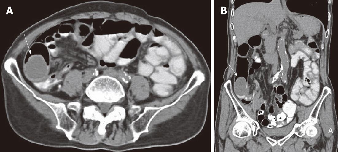
Figure 6 A 79-year-old male with ileum malignant fibrous histiocytoma, plain computed tomography scan abdomen.
A, B: Axial (A) and coronal view (B) show intraluminal and intramural soft tissue mass of density 34 HU in the ilio-cecal region (arrows) without significant areas of necrosis. Note the oral contrast stagnation in the proximal loop of the small intestine and dilatation of the ascending colon.
- Citation: Karki B, Xu YK, Wu YK, Zhang WW. Primary malignant fibrous histiocytoma of the abdominal cavity: CT findings and pathological correlation. World J Radiol 2012; 4(4): 151-158
- URL: https://www.wjgnet.com/1949-8470/full/v4/i4/151.htm
- DOI: https://dx.doi.org/10.4329/wjr.v4.i4.151