Copyright
©2012 Baishideng Publishing Group Co.
World J Radiol. Apr 28, 2012; 4(4): 141-150
Published online Apr 28, 2012. doi: 10.4329/wjr.v4.i4.141
Published online Apr 28, 2012. doi: 10.4329/wjr.v4.i4.141
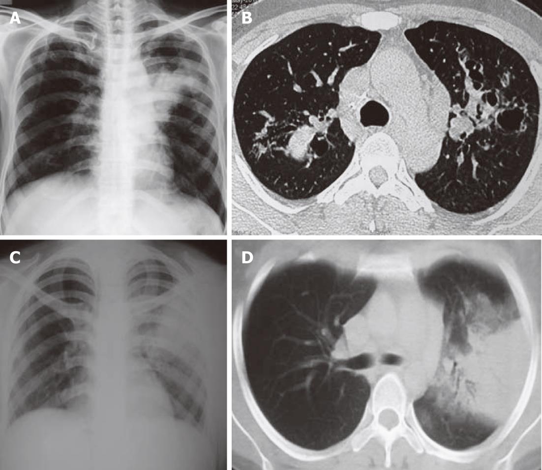
Figure 2 Chest radiograph.
A, B: Chest radiograph (right panel) demonstrating consolidation in the left upper zone. Corresponding high resolution computed tomography of the chest (left panel) shows mucus-filled dilated bronchi; C, D: Chest radiograph (right panel) demonstrating consolidation in left upper zone. Computed tomography chest (left panel) shows typical consolidation with air bronchograms.
- Citation: Agarwal R, Khan A, Garg M, Aggarwal AN, Gupta D. Chest radiographic and computed tomographic manifestations in allergic bronchopulmonary aspergillosis. World J Radiol 2012; 4(4): 141-150
- URL: https://www.wjgnet.com/1949-8470/full/v4/i4/141.htm
- DOI: https://dx.doi.org/10.4329/wjr.v4.i4.141


