Copyright
©2012 Baishideng Publishing Group Co.
World J Radiol. Apr 28, 2012; 4(4): 128-134
Published online Apr 28, 2012. doi: 10.4329/wjr.v4.i4.128
Published online Apr 28, 2012. doi: 10.4329/wjr.v4.i4.128
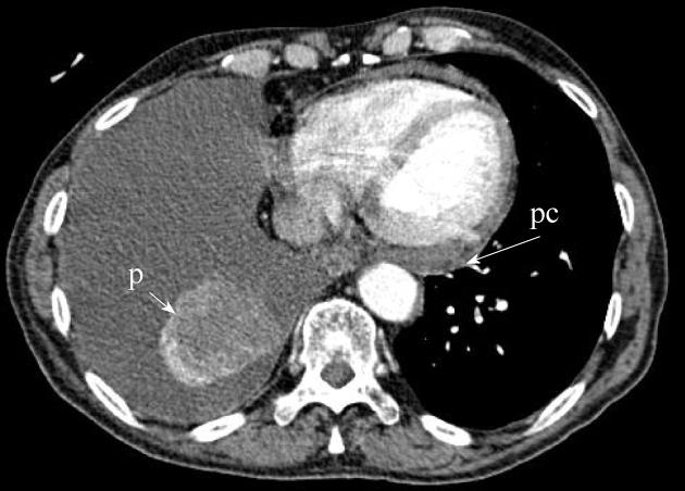
Figure 7 A patient with a necrotic mass in the right lower lobe (short arrow).
As seen in this axial contrast enhanced computed tomography, there is pleural (p) and pericardial (pc) effusions which were confirmed to be malignant. This will be re-classified from T4 to M1a indicating worse estimated prognosis.
- Citation: Mirsadraee S, Oswal D, Alizadeh Y, Caulo A, Beek EJV. The 7th lung cancer TNM classification and staging system: Review of the changes and implications. World J Radiol 2012; 4(4): 128-134
- URL: https://www.wjgnet.com/1949-8470/full/v4/i4/128.htm
- DOI: https://dx.doi.org/10.4329/wjr.v4.i4.128


