Copyright
©2012 Baishideng Publishing Group Co.
World J Radiol. Mar 28, 2012; 4(3): 102-108
Published online Mar 28, 2012. doi: 10.4329/wjr.v4.i3.102
Published online Mar 28, 2012. doi: 10.4329/wjr.v4.i3.102
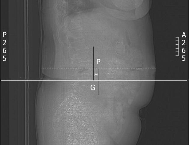
Figure 1 Calculation of off-centering distance.
First, the gantry isocenter (G) was determined as the midpoint of the entire image (thick line). Next, maximal anterior-posterior diameter of the patient (broken line) was measured. Lastly, distance (asterix) between the midpoints of maximal patient diameter (P) and gantry isocenter (G) was measured to obtain the off-centering distance.
- Citation: Kim MS, Singh S, Halpern E, Saini S, Kalra MK. Ablation margin assessment of liver tumors with intravenous contrast-enhanced C-arm computed tomography. World J Radiol 2012; 4(3): 102-108
- URL: https://www.wjgnet.com/1949-8470/full/v4/i3/102.htm
- DOI: https://dx.doi.org/10.4329/wjr.v4.i3.102


