Copyright
©2012 Baishideng Publishing Group Co.
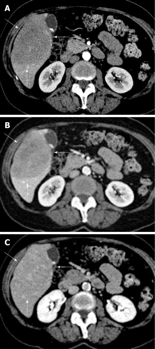
Figure 1 Radiological depiction of the liver lesion.
Arterial (A), portal (B) and equilibrium phase (C) computed tomography scan with a large (10 cm × 8 cm × 7.5 cm) hypovascular lesion in segments V and VI of the liver with some inhomogeneity and without calcifications (lesion indicated by 3 white arrows).
- Citation: Steller EJ, Leeuwen MSV, Hillegersberg RV, Schipper ME, Rinkes IHB, Molenaar IQ. Primary lymphoma of the liver - A complex diagnosis. World J Radiol 2012; 4(2): 53-57
- URL: https://www.wjgnet.com/1949-8470/full/v4/i2/53.htm
- DOI: https://dx.doi.org/10.4329/wjr.v4.i2.53


