Copyright
©2012 Baishideng Publishing Group Co.
World J Radiol. Oct 28, 2012; 4(10): 439-442
Published online Oct 28, 2012. doi: 10.4329/wjr.v4.i10.439
Published online Oct 28, 2012. doi: 10.4329/wjr.v4.i10.439
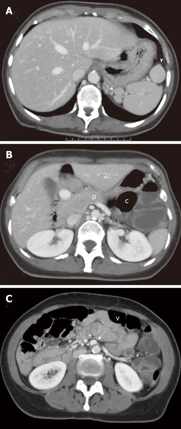
Figure 4 Multi-detector contrast-enhanced computed tomography.
Transverse images at the level of the upper (A and B) and the middle abdomen (C) are shown. Retrospective reading of computed tomography images revealed multiple splenic nodules (arrowheads) in the left sub-phrenic space (A) as well as a truncated appearance of the pancreatic body (p) (B) and an inverted anatomic relationship between the mesenteric superior artery (a) and vein (v) (C). c: Colon.
- Citation: Camera L, Calabrese M, Mainenti PP, Masone S, Vecchio WD, Persico G, Salvatore M. Volvulus of the ascending colon in a non-rotated midgut: Plain film and MDCT findings. World J Radiol 2012; 4(10): 439-442
- URL: https://www.wjgnet.com/1949-8470/full/v4/i10/439.htm
- DOI: https://dx.doi.org/10.4329/wjr.v4.i10.439


