Copyright
©2012 Baishideng Publishing Group Co.
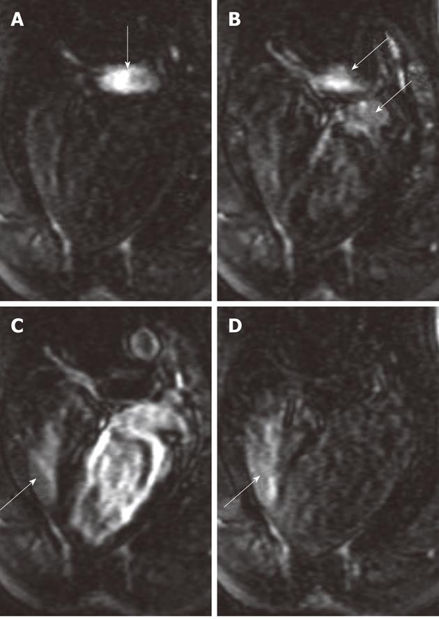
Figure 7 Bolus administration of gadolinium-chelate demonstrates atrial septal defect on cardiac magnetic resonance four-chamber view images.
At the time of administration of the contrast medium, the right atrium was enhanced (A, arrow). Images acquired 1-2 s later show the contrast medium in both the pulmonary artery and left atrium (B, arrows) and subsequently in both right (arrow) and left ventricles (C). As a sign of an intracardiac left-to-right shunt, enhancement of the right ventricle is detected simultaneously with enhancement of the left ventricle. The re-enhancement of the right ventricle (D, arrow) as a result of recirculation of the contrast medium was acquired 12 s after image A.
- Citation: Saeed M, Wilson M. Value of MR contrast media in image-guided body interventions. World J Radiol 2012; 4(1): 1-12
- URL: https://www.wjgnet.com/1949-8470/full/v4/i1/1.htm
- DOI: https://dx.doi.org/10.4329/wjr.v4.i1.1


