Copyright
©2011 Baishideng Publishing Group Co.
World J Radiol. Sep 28, 2011; 3(9): 224-232
Published online Sep 28, 2011. doi: 10.4329/wjr.v3.i9.224
Published online Sep 28, 2011. doi: 10.4329/wjr.v3.i9.224
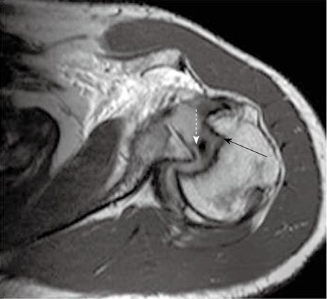
Figure 18 Reverse Hill-Sachs and reverse Bankart lesion in a case of posterior instability.
T1-weighted TSE axial magnetic resonance image reveals hemarthrosis, posterior glenohumeral dislocation and reverse Hill-Sachs lesion (straight arrow). There is associated posterior labral tear (reverse Bankart lesion), shown with a dashed arrow.
- Citation: Jana M, Gamanagatti S. Magnetic resonance imaging in glenohumeral instability. World J Radiol 2011; 3(9): 224-232
- URL: https://www.wjgnet.com/1949-8470/full/v3/i9/224.htm
- DOI: https://dx.doi.org/10.4329/wjr.v3.i9.224


