Copyright
©2011 Baishideng Publishing Group Co.
World J Radiol. Sep 28, 2011; 3(9): 215-223
Published online Sep 28, 2011. doi: 10.4329/wjr.v3.i9.215
Published online Sep 28, 2011. doi: 10.4329/wjr.v3.i9.215
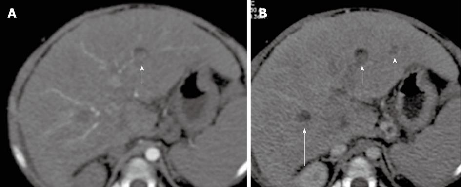
Figure 14 A 20-mo-old female without previous Kasai operation.
Multi-detector computed tomography: In S3, nodule (short arrow) that shows in anterior portion a mild amount of fat. The nodule appears slightly hyperdense in arterial phase (A) and hypoattenuating in portal/venous phase; B: Regenerative nodule was found at liver explant. Of note: other hypoattenuating regenerative nodules in S6 and S3 (long arrows).
- Citation: Miraglia R, Caruso S, Maruzzelli L, Spada M, Riva S, Sciveres M, Luca A. MDCT, MR and interventional radiology in biliary atresia candidates for liver transplantation. World J Radiol 2011; 3(9): 215-223
- URL: https://www.wjgnet.com/1949-8470/full/v3/i9/215.htm
- DOI: https://dx.doi.org/10.4329/wjr.v3.i9.215


