Copyright
©2011 Baishideng Publishing Group Co.
World J Radiol. Sep 28, 2011; 3(9): 215-223
Published online Sep 28, 2011. doi: 10.4329/wjr.v3.i9.215
Published online Sep 28, 2011. doi: 10.4329/wjr.v3.i9.215
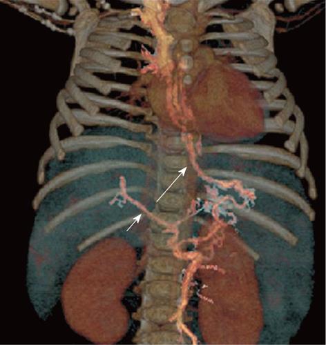
Figure 8 A 8-mo-old female child post-Kasai.
Multi-detector computed tomography: volume rendering reconstruction shows small main portal vein (short arrow), patent coronary vein (long arrow) with filling of esophageal varices.
- Citation: Miraglia R, Caruso S, Maruzzelli L, Spada M, Riva S, Sciveres M, Luca A. MDCT, MR and interventional radiology in biliary atresia candidates for liver transplantation. World J Radiol 2011; 3(9): 215-223
- URL: https://www.wjgnet.com/1949-8470/full/v3/i9/215.htm
- DOI: https://dx.doi.org/10.4329/wjr.v3.i9.215


