Copyright
©2011 Baishideng Publishing Group Co.
World J Radiol. Aug 28, 2011; 3(8): 199-204
Published online Aug 28, 2011. doi: 10.4329/wjr.v3.i8.199
Published online Aug 28, 2011. doi: 10.4329/wjr.v3.i8.199
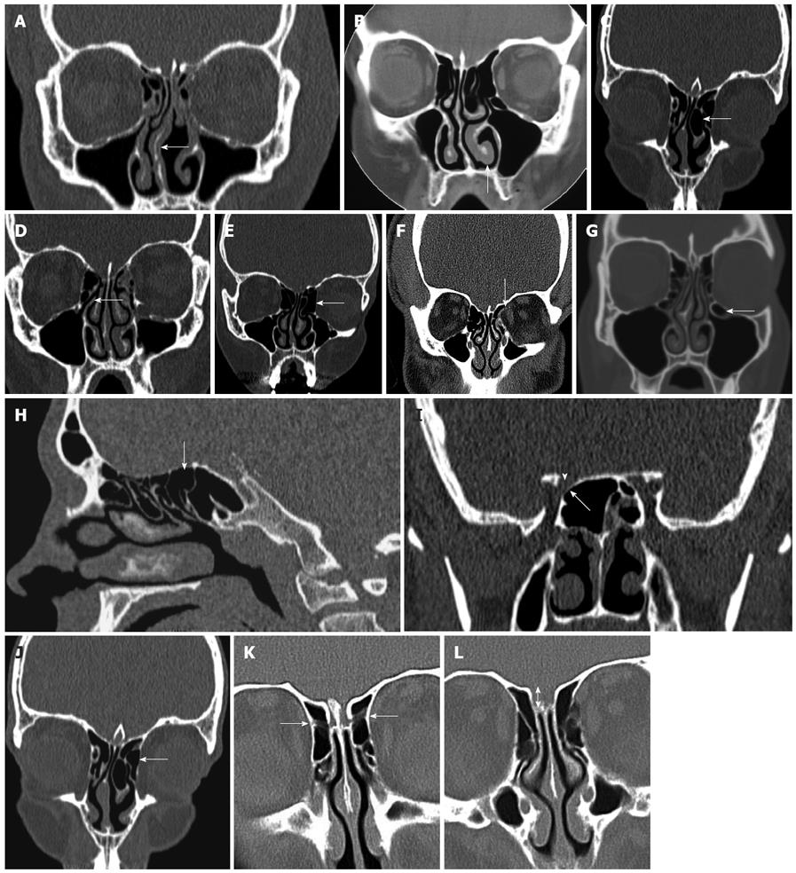
Figure 2 Coronal computed tomography reformat.
A: The paranasal sinuses demonstrating a deviated septum to the right side (arrow); B: Mucosal thickening of the inferior turbinate on the left side (arrow); C: A pneumatized left middle turbinate (arrow); D: The uncinate process (arrow); E: The ethmoid bulla adjacent to the left orbit (arrow), note its extent to the floor of the anterior cranial fossa cranially and lamina papyracea laterally; F: A dehiscent lamina papyracea on the left side (arrow); G: An infra-orbital cell (Haller cell) (arrow); H: The posterior ethmoid cell (arrow) directly anterior to the sphenoid sinus; I: At level of sphenoid sinuses, thinning of the bony in the superolateral right sphenoid sinus adjacent to the right optic nerve (arrow head); J: The agger nasi cell (arrow) which lies inferior to the frontal recess and lateral to the middle turbinate; K: The anterior ethmoidal artery (arrow) traversing the anterior ethmoid air cells, which is susceptible to injury during functional endoscopic sinus surgery; L: A type III olfactory fossa (arrow).
- Citation: Cashman EC, MacMahon PJ, Smyth D. Computed tomography scans of paranasal sinuses before functional endoscopic sinus surgery. World J Radiol 2011; 3(8): 199-204
- URL: https://www.wjgnet.com/1949-8470/full/v3/i8/199.htm
- DOI: https://dx.doi.org/10.4329/wjr.v3.i8.199


