Copyright
©2011 Baishideng Publishing Group Co.
World J Radiol. Dec 28, 2011; 3(12): 279-288
Published online Dec 28, 2011. doi: 10.4329/wjr.v3.i12.279
Published online Dec 28, 2011. doi: 10.4329/wjr.v3.i12.279
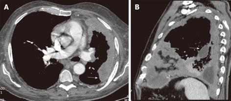
Figure 9 Malignant mesothelioma in a 60-year-old male with progressive chest pain.
Contrast-enhanced computed tomography, axial (A) and sagittal (B) images show an irregular and slightly lobulated soft tissue density mass extensively involving the pleural surface of the left lung including the major fissure (arrows).
- Citation: Restrepo CS, Chen MM, Martinez-Jimenez S, Carrillo J, Restrepo C. Chest neoplasms with infectious etiologies. World J Radiol 2011; 3(12): 279-288
- URL: https://www.wjgnet.com/1949-8470/full/v3/i12/279.htm
- DOI: https://dx.doi.org/10.4329/wjr.v3.i12.279


